Ear Anatomy 3 Times Magnification Divided into 2 Parts Ear and Ear Inner Ear Hearing Otolaryngology
Availability:
In stock
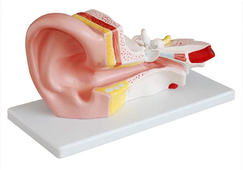
Explore the human ear in stunning detail with our 3x magnified, 2-part dissectible ear anatomy model. Perfect for otolaryngology and medical education, this model provides an accurate, clear view of the outer, middle, and inner ear structures.
Ear Anatomy Model: 3x Magnification, 2-Part Dissectible
Our Ear Anatomy Model provides an unparalleled, in-depth look at the intricate structures of the human ear. Magnified three times to life size, this model is an essential teaching tool for medical students, otolaryngology professionals, and educators. It’s designed to make complex anatomy easy to understand, both in the classroom and the clinic.
Unmatched Detail and Clarity
This model is meticulously crafted to showcase the three main sections of the ear: the outer ear, middle ear, and inner ear.
-
Outer Ear: Features the auricle (pinna) and the external auditory canal.
-
Middle Ear: Clearly displays the tympanic membrane (eardrum) and the three auditory ossicles: the malleus, incus, and stapes.
-
Inner Ear: The most complex part, with the cochlea, semicircular canals, and the auditory and vestibular nerves, is shown in stunning detail.
Key Features
-
2-Part Dissectible Design: The model is intelligently divided into two parts to allow for a focused examination of the inner ear. One section detaches, giving you a clear view of the complex labyrinth, including the cochlea for hearing and the semicircular canals for balance.
-
High-Quality Construction: Made from durable, medical-grade PVC, this model is built to withstand frequent use in a lecture hall or doctor’s office. The vibrant, hand-painted details are accurate and fade-resistant.
-
Otolaryngology & Patient Education: Ideal for otolaryngologists, audiologists, and general practitioners to explain conditions like hearing loss, vertigo, and middle ear infections to patients. Its large size and clear labelling make it a perfect aid for patient communication.
This anatomical model simplifies the study of hearing and balance, transforming a challenging subject into a visually engaging and memorable learning experience.
Related products
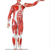
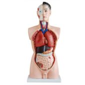
male muscle figure life size 170 cm with internal organs
Life-size Male Muscle Figure 170 cm with removable internal organs. High-quality anatomical model for medical training, anatomy education, and clinical demonstrations.

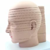
deluxe model of human disc head horizontally sliced
The Deluxe Model of Human Disc Head Horizontally Sliced offers detailed cross-sectional anatomy of the human head. Ideal for medical training, teaching, and anatomical study.
Reviews
There are no reviews yet.