eye with orbit and muscles model
Availability:
In stock
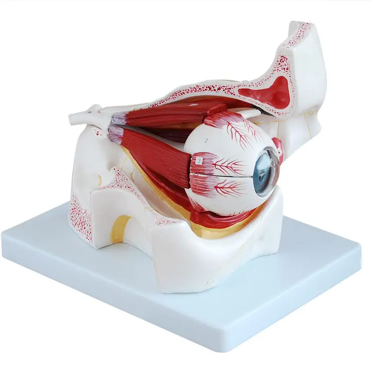
Eye with Orbit and Muscles Model: A detailed anatomical model of the human eye and its six extraocular muscles within the bony orbit. Perfect for medical students and educators to study eye movement and anatomy.
Gain a deeper understanding of the human eye with our highly detailed Eye with Orbit and Muscles Model. This professional-grade anatomical model provides a realistic, three-dimensional representation of the eyeball, its complex network of surrounding muscles, and its bony socket (the orbit). It is an invaluable educational tool for students, ophthalmologists, and medical professionals.
This model is meticulously crafted to showcase the intricate relationship between the eye and its supporting structures. Key features include:
-
The Eyeball: A life-size or enlarged depiction of the eyeball, often dissectible into multiple parts to reveal the internal structures such as the cornea, lens, retina, and optic nerve.
-
Extraocular Muscles: All six extraocular muscles—the superior, inferior, medial, and lateral rectus muscles, as well as the superior and inferior oblique muscles—are accurately represented. Their origins on the bony orbit and their insertions on the sclera are clearly shown, allowing for a clear demonstration of eye movement and function.
-
The Bony Orbit: The model features a detailed bony socket, or orbit, formed by a mosaic of several skull bones. This enables a comprehensive examination of how the eye is protected and supported within the skull.
-
Durable and High-Quality Construction: Made from washable, durable PVC, this model is designed for long-lasting use in classrooms and labs. The hand-painted, vibrant colors enhance visual learning and make complex anatomy easy to identify.
With its dissectible components and precise anatomical detail, this model is an essential resource for exploring the mechanics of vision, the causes of eye disorders, and the intricate workings of the visual system. It’s the perfect way to bring the complex anatomy of the eye to life.
Related products
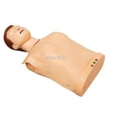
half body electronic cpr training manikin with indicator
₨0.00
Enhance CPR training with our Half Body Electronic CPR Training Manikin featuring a built-in indicator. Simulate realistic scenarios and track progress efficiently. Elevate your life-saving skills with this advanced training tool. Shop now for a more innovative approach to CPR education.
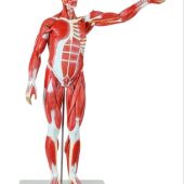
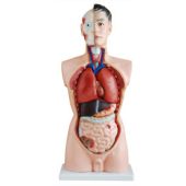
male muscle figure life size 170 cm with internal organs
Life-size Male Muscle Figure 170 cm with removable internal organs. High-quality anatomical model for medical training, anatomy education, and clinical demonstrations.
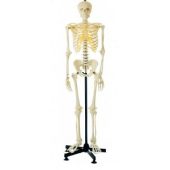
human skeleton life-size male 180CM tall
This Classic Human Skeleton has been the standard of excellence and quality in all major Medical Colleges, Hospitals, Universities, School and Laboratories.

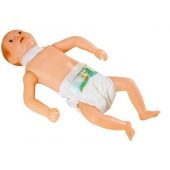
advance infant cpr training manikin soft
Realistic Advance Infant CPR Training Manikin (Soft) for effective infant resuscitation practice. Ideal for CPR courses, nursing schools, and emergency medical training.
infant tracheostomy care simulator soft
Train safely with the Infant Tracheostomy Care Simulator Soft, a realistic model for practicing pediatric tracheostomy care, suctioning, and tube management.
Reviews
There are no reviews yet.